Blood Vessels Labeled Diagram - Blood vessels - Veins in arm & shoulder - PurposeGames : Jun 17, 2021 · let’s put into words the heart blood flow diagram:
byAdmin•
0
Blood Vessels Labeled Diagram - Blood vessels - Veins in arm & shoulder - PurposeGames : Jun 17, 2021 · let's put into words the heart blood flow diagram:. Write no more than two words from the passage for each answer. Blood from both atria goes into the ventricle and then is pumped into the arteries, which are blood vessels that carry blood away from the heart. The recommended location for blood collection on a newborn baby or infant is the heel. It is an antique print to be used for various purposes. Includes anatomy of the femur quiz.
Jun 17, 2021 · let's put into words the heart blood flow diagram: Blood leaves the left ventricle of the heart through the aortic semilunar valve and enters the aorta. The urinary system consists of the frog's kidneys, ureters, bladder, and. Label the diagram using the descriptions and bold words. The recommended location for blood collection on a newborn baby or infant is the heel.
Blood Vessels: Arteries, Capillaries & More - Video ... from study.com The pulmonary circuit is responsible for exchanging blood between the heart and lungs for oxygenation, while the systemic circuit directs blood to the other tissues of the body. Write no more than two words from the passage for each answer. Mar 29, 2021 · femur bone anatomy made easy using a labeled diagram of the main parts of the thigh bone along with their location. Trace the flow of blood using arrows. Prewarming the infant's heel (42° c for 3 to 5 minutes) is important to increase the flow of blood for collection. Blood leaves the left ventricle of the heart through the aortic semilunar valve and enters the aorta. Blood from both atria goes into the ventricle and then is pumped into the arteries, which are blood vessels that carry blood away from the heart. The recommended location for blood collection on a newborn baby or infant is the heel.
This is an illustration from an old medical book which not only shows the heart diagram but also the concerned blood vessels.
Jun 17, 2021 · let's put into words the heart blood flow diagram: Jul 27, 2021 · comprised of the heart, blood vessels and the blood itself, it is divided into two loops which both begin in the heart. The aorta has a visible arch with vessels that lead to the head before the artery descends into the rat's thoracic cavity. The pulmonary circuit is responsible for exchanging blood between the heart and lungs for oxygenation, while the systemic circuit directs blood to the other tissues of the body. This is an illustration from an old medical book which not only shows the heart diagram but also the concerned blood vessels. Veins from different parts of the body enter the right and left atria. Prewarming the infant's heel (42° c for 3 to 5 minutes) is important to increase the flow of blood for collection. Mar 29, 2021 · femur bone anatomy made easy using a labeled diagram of the main parts of the thigh bone along with their location. The urinary system consists of the frog's kidneys, ureters, bladder, and. Blood from both atria goes into the ventricle and then is pumped into the arteries, which are blood vessels that carry blood away from the heart. It is an antique print to be used for various purposes. High blood pressure is classified as primary (essential) hypertension or secondary hypertension. Fractures to the femur and hip bone can occur and knowing the anatomy will help with management.
High blood pressure is classified as primary (essential) hypertension or secondary hypertension. The diagram below indicates the proper area to use for heel punctures for blood collection. The pulmonary circuit is responsible for exchanging blood between the heart and lungs for oxygenation, while the systemic circuit directs blood to the other tissues of the body. Blood from both atria goes into the ventricle and then is pumped into the arteries, which are blood vessels that carry blood away from the heart. Includes anatomy of the femur quiz.
Home Dialysis Central from www.homedialysis.org The right atrium receives deoxygenated blood from the superior and inferior venae cavae and coronary sinus the right atrium contracts pushing blood through the right atrioventricular valve into the right ventricle. The pulmonary circuit is responsible for exchanging blood between the heart and lungs for oxygenation, while the systemic circuit directs blood to the other tissues of the body. Fractures to the femur and hip bone can occur and knowing the anatomy will help with management. Mar 29, 2021 · femur bone anatomy made easy using a labeled diagram of the main parts of the thigh bone along with their location. Veins from different parts of the body enter the right and left atria. Label the diagram using the descriptions and bold words. The recommended location for blood collection on a newborn baby or infant is the heel. Jul 27, 2021 · comprised of the heart, blood vessels and the blood itself, it is divided into two loops which both begin in the heart.
Fractures to the femur and hip bone can occur and knowing the anatomy will help with management.
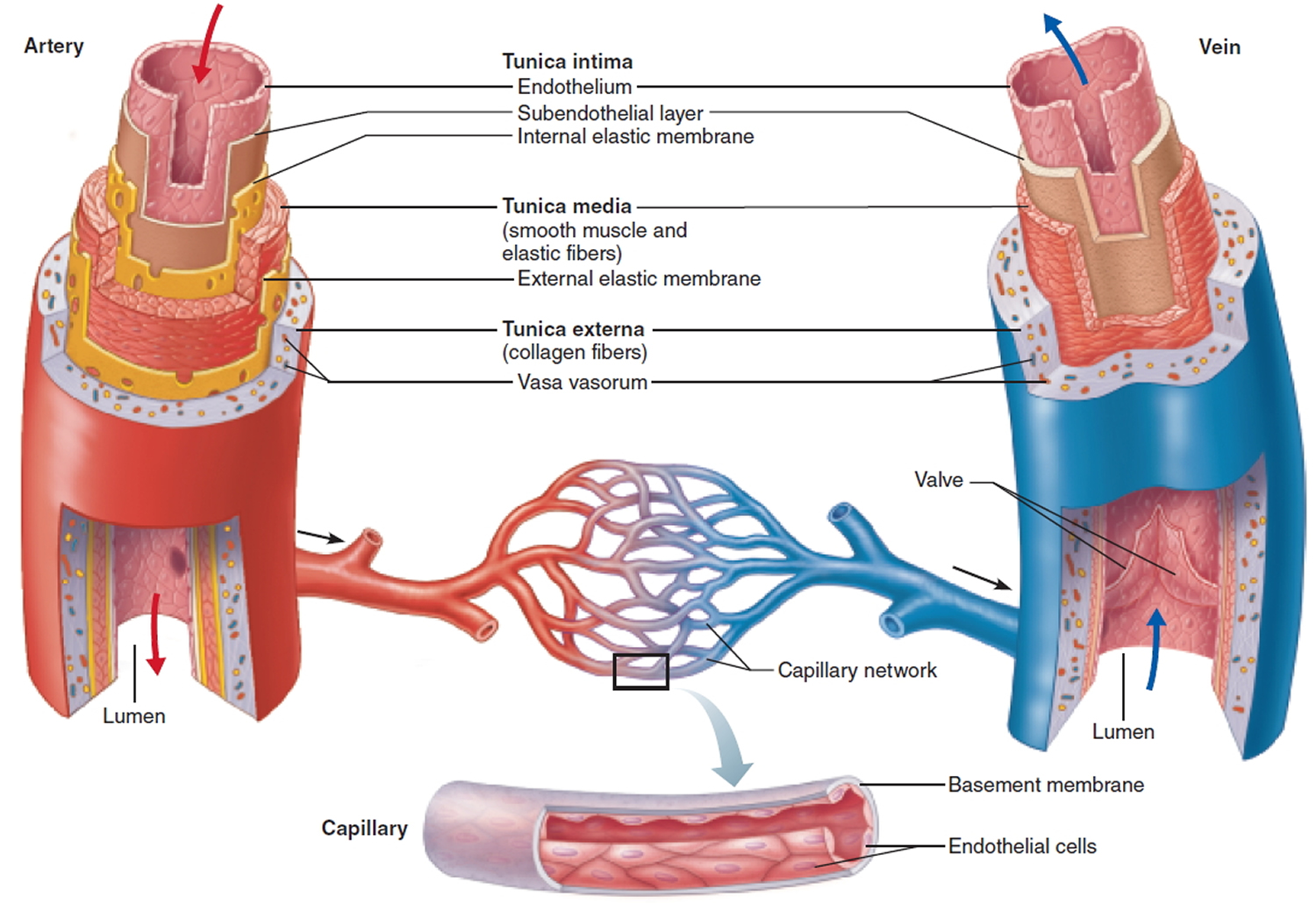
Prewarming the infant's heel (42° c for 3 to 5 minutes) is important to increase the flow of blood for collection. The urinary system consists of the frog's kidneys, ureters, bladder, and. Jun 17, 2021 · let's put into words the heart blood flow diagram: Trace the flow of blood using arrows. Blood is carried to the heart in vessels called veins. Jul 27, 2021 · comprised of the heart, blood vessels and the blood itself, it is divided into two loops which both begin in the heart. The recommended location for blood collection on a newborn baby or infant is the heel. Write no more than two words from the passage for each answer. Veins from different parts of the body enter the right and left atria. This is an illustration from an old medical book which not only shows the heart diagram but also the concerned blood vessels. The word vascular, meaning relating to the blood vessels, is derived from the latin vas, meaning vessel. Fractures to the femur and hip bone can occur and knowing the anatomy will help with management. The embryo develops from this disk, and gradually sends blood vessels into the yolk to use it for nutrition as the embryo develops.
Prewarming the infant's heel (42° c for 3 to 5 minutes) is important to increase the flow of blood for collection. Label the diagram using the descriptions and bold words. Mar 29, 2021 · femur bone anatomy made easy using a labeled diagram of the main parts of the thigh bone along with their location. High blood pressure is classified as primary (essential) hypertension or secondary hypertension. Write no more than two words from the passage for each answer.
Blood Vessels Types - Layers of Blood Vessels - Carry ... from healthjade.com The diagram below indicates the proper area to use for heel punctures for blood collection. The urinary system consists of the frog's kidneys, ureters, bladder, and. This is an illustration from an old medical book which not only shows the heart diagram but also the concerned blood vessels. The recommended location for blood collection on a newborn baby or infant is the heel. Mar 29, 2021 · femur bone anatomy made easy using a labeled diagram of the main parts of the thigh bone along with their location. Blood leaves the left ventricle of the heart through the aortic semilunar valve and enters the aorta. Label the diagram using the descriptions and bold words. Prewarming the infant's heel (42° c for 3 to 5 minutes) is important to increase the flow of blood for collection.
Includes anatomy of the femur quiz.
Blood leaves the left ventricle of the heart through the aortic semilunar valve and enters the aorta. The right atrium receives deoxygenated blood from the superior and inferior venae cavae and coronary sinus the right atrium contracts pushing blood through the right atrioventricular valve into the right ventricle. High blood pressure is classified as primary (essential) hypertension or secondary hypertension. The word vascular, meaning relating to the blood vessels, is derived from the latin vas, meaning vessel. Jul 27, 2021 · comprised of the heart, blood vessels and the blood itself, it is divided into two loops which both begin in the heart. Trace the flow of blood using arrows. Blood from both atria goes into the ventricle and then is pumped into the arteries, which are blood vessels that carry blood away from the heart. Mar 29, 2021 · femur bone anatomy made easy using a labeled diagram of the main parts of the thigh bone along with their location. Includes anatomy of the femur quiz. The recommended location for blood collection on a newborn baby or infant is the heel. The diagram below indicates the proper area to use for heel punctures for blood collection. This is an illustration from an old medical book which not only shows the heart diagram but also the concerned blood vessels. Blood is carried to the heart in vessels called veins.
Fractures to the femur and hip bone can occur and knowing the anatomy will help with management blood vessels labeled. The recommended location for blood collection on a newborn baby or infant is the heel.